Time Signal Intensity Curve Breast Mri
Benignmalignant overlap exists on time intensity curve tic of dynamic gadolinium enhanced breast mri. The tic is obtained by repeated mri scans after the injection of contrast agent.
Http Egyptianjournal Xyz 53 20 Pdf
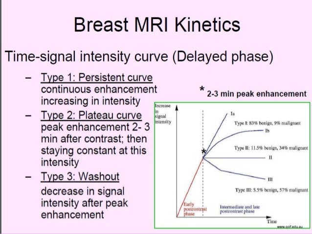
Progressive enhancement pattern typically shows a continuous increase in signal intensity throughout time.

Time signal intensity curve breast mri. Following administration of gadolinium there can be three possible enhancement kinetic curves for a lesion on breast mri these are also applied in other organs such as prostate mri. The shape of the timesignal intensity curve is an important criterion in differentiating benign and malignant enhancing lesions in dynamic breast mr imaging. The tic is obtained by repeated mri scans after the injection of contrast agent.
Generally speaking invasive cancer in the breast will show a more than 70 increase in signal intensity over baseline within the first 60 90 seconds because of large vessels in the tumor. This study presents a new method for tic generation to decrease overlap and improve accuracy. In one study further evaluation with mri was shown to find additional foci of cancer in 13 of patients diagnosed with ilc at biopsy who were anticipating breast conservation therapy.
These are sometimes termed the kuhl enhancement curves. A qualified tic usually takes 12 minutes to complete the scans. Adding to the diagnostic challenges is the propensity for ilc to be bilateral and multicentric 20 30 of the time.
In breast mri by plotting the signal intensity values over time after the administration of contrast material. A type iii time course is a strong indicator of malignancy and is independent of other criteria. The time signal intensity curve tic from dynamic contrast enhanced magnetic resonance imaging dce mri reflects the hemodynamic features of a specific lesion.
This marked increase in signal intensity is followed by a wash out phase. A qualified tic usually takes 12 minutes to complete the scans. The time intensity curve tool allows you to visualize the lesions enhancement behavior eg.
The time signal intensity curve tic from dynamic contrast enhanced magnetic resonance imaging dce mri reflects the hemodynamic features of a specific lesion.
Presentation1 Pptx Radiological Imaging Of Beign Breast Diseases

Breast Cancer Radiology Key
Imaging Of Soft Tissue Sarcomas Patel Chinese Clinical Oncology

Vbl


Automated Volumetric Radiomic Analysis Of Breast Cancer
Time Intensity Curves Tic

Typical Curve Shapes For Qualitative Dce Breast Mri Analysis Si
Pdf Histopathological Correlation Of Time Signal Intensity Curve

The Radiology Assistant Breast Mri